Jones Criteria Explained: Diagnosing Acute Rheumatic Fever with Confidence

Most clinicians learn the diagnostic rules for acute rheumatic fever during training, then encounter a case that refuses to fit neatly into any category. The Jones Criteria exist precisely to prevent that diagnostic drift. They provide a disciplined, evidence-based framework that — when applied correctly — allows clinicians to confirm streptococcal exposure, map clinical features against validated thresholds, and reach a defensible diagnosis with both speed and precision. The real contest in acute rheumatic fever is not the diagnosis itself — it is preventing the progression to rheumatic heart disease that follows a missed or delayed one. At Moolchand Hospital's Cardiology and Paediatric Medicine Department, the Jones Criteria are applied systematically alongside early echocardiography and structured secondary prophylaxis planning. This guide explains exactly how.
The Structure of the Jones Criteria
The Jones Criteria divide diagnostic features into two tiers — major and minor — and require these to be combined with objective evidence of a recent group A streptococcal infection. Understanding the distinction between tiers, and between risk populations, is the foundation of accurate application.
Major Criteria in Low-Risk Populations
In standard, low-incidence settings, five major manifestations are recognised. Each carries significant weight because of its association with cardiac involvement and long-term rheumatic heart disease risk.
1. Carditis — either clinically evident or subclinical valvulitis meeting echocardiographic Doppler standards. This is the most consequential major criterion because it directly predicts long-term valve damage.
2. Migratory polyarthritis — large joint inflammation that characteristically moves from joint to joint. Knees, ankles, elbows, and wrists are most commonly involved. The migratory pattern is a specific and diagnostically valuable feature.
3. Sydenham Chorea — involuntary, purposeless, jerky movements caused by basal ganglia inflammation. Chorea may arrive weeks to months after the initial infection and is sometimes the only presenting feature, which the Jones Criteria explicitly accommodate.
4. Erythema marginatum — an evanescent, non-pruritic rash with characteristic serpiginous, advancing edges, typically appearing on the trunk and proximal limbs.
5. Subcutaneous nodules — firm, painless lumps distributed over bony prominences and extensor tendon surfaces. These are now less common but clinically distinctive when present.
In everyday practice, arthritis and carditis account for the majority of presentations. Chorea may arrive late and in isolation. The Jones Criteria are designed to accept this timing asymmetry to protect against false negatives.
Major Criteria in High-Risk Populations
Where disease burden is higher — in settings characterised by overcrowding, limited healthcare access, or known endemic rheumatic heart disease — the Jones Criteria broaden the joint criteria to reflect observed real-world phenotypes. In these populations, monoarthritis or polyarthralgia (joint pain without swelling) may qualify as a major criterion rather than a minor one. This adjustment reflects the fact that severe systemic inflammation in high-incidence settings can concentrate in a single joint — a signal that should be treated seriously rather than discounted because it does not match the textbook polyarticular pattern. Carditis, chorea, erythema marginatum, and subcutaneous nodules carry the same definitions across both risk groups.
Minor Criteria: Calibrated to Context
Minor criteria provide supporting evidence that strengthens the diagnostic picture when combined with a major criterion. Crucially, the Jones Criteria specify different thresholds for low-risk and high-risk populations — a design feature that keeps the framework calibrated to clinical reality rather than abstract norms.
|
Minor
Criterion |
Low-Risk
Threshold |
High-Risk
Threshold |
|
Arthralgia |
Polyarthralgia |
Monoarthralgia |
|
Fever |
≥ 38.5°C |
≥ 38.0°C |
|
ESR |
≥ 60 mm/hour |
≥ 30 mm/hour |
|
CRP |
≥ 3.0 mg/dL |
≥ 3.0 mg/dL |
|
PR interval |
Prolonged for age |
Prolonged for age |
When the risk population is uncertain, applying high-risk thresholds is always the safer choice. The Jones Criteria are designed to err towards sensitivity where the consequences of a missed case — progressive valve damage — are significantly worse than the consequences of an over-call.
Subclinical Carditis on Echocardiography
One of the most important advances in acute rheumatic fever diagnosis over the past two decades is the formal recognition of subclinical carditis as a major Jones criterion. Subclinical carditis is present on Doppler echocardiography but produces no audible murmur on clinical examination. In high-incidence settings, it is common — and catching it early substantially changes prognosis and prophylaxis planning.
For subclinical carditis to qualify as a major criterion under the Jones Criteria, specific standards must be met:
- Pathological mitral regurgitation: regurgitant jet visible in at least two imaging views, meeting defined velocity and jet-length criteria
- Pathological aortic regurgitation: equivalent Doppler standards in at least two views
- Valve morphology: leaflet thickening, restricted motion, chordal changes, or prolapse consistent with rheumatic valvulitis — not physiological trivial regurgitation
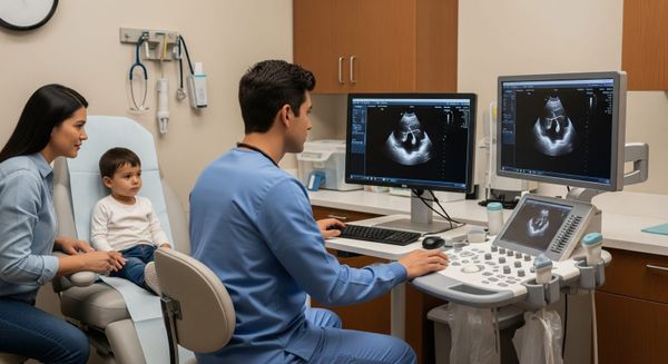
The clinical implication is direct: echocardiography should be requested for every suspected case of acute rheumatic fever, not reserved for those with audible murmurs. The Cardiac Imaging and Echocardiography team at Moolchand Hospital performs dedicated Doppler echocardiographic assessment as an integrated part of the rheumatic fever diagnostic pathway, ensuring subclinical carditis is identified at the earliest possible stage.
Confirming Streptococcal Exposure
The Jones Criteria require objective evidence of a recent group A streptococcal infection in most presentations. This can be established through any of the following:
- Positive throat culture or rapid antigen detection test for group A streptococcus
- Elevated or rising streptococcal antibody titres — antistreptolysin O (ASO) or anti-DNase B — on paired samples
- A documented episode of scarlet fever in the preceding weeks
Two recognised exceptions apply. In Sydenham chorea, the latency between infection and the neurological presentation may be 1–6 months — long enough for both throat tests and antibody titres to normalise. In indolent carditis with late valve involvement, recent infection evidence may similarly be absent. The Jones Criteria explicitly permit diagnosis in both these scenarios on the basis of clinical and echocardiographic findings alone, with secondary prophylaxis then mandatory. The Pathology and Diagnostic Laboratory at Moolchand Hospital provides same-day results for rapid antigen testing, ASO titres, anti-DNase B, CRP, and ESR — the complete panel required for Jones Criteria confirmation.
Diagnostic Combinations: What Is Required
For a confirmed initial episode of acute rheumatic fever, the Jones Criteria require one of the following combinations, always alongside proof of streptococcal infection:
- Two major criteria, or
- One major plus two minor criteria
For a recurrent episode in a patient with previously documented rheumatic carditis, the threshold is appropriately lower — reflecting the higher probability of genuine recurrence in this group. Two major, or one major plus two minor, or three minor criteria are all acceptable with confirmed streptococcal exposure.
For chorea and indolent carditis, diagnosis proceeds on clinical and echocardiographic grounds without requiring serological proof of infection. In every case, the exact criteria combination met should be explicitly documented in the clinical record. The Jones Criteria serve as both a clinical and a medicolegal framework, and clarity in documentation protects patients and clinicians alike.
Differentiating Acute Rheumatic Fever from Mimics
Post-Streptococcal Reactive Arthritis
Post-streptococcal reactive arthritis (PSRA) is the most clinically significant differential to separate from genuine acute rheumatic fever. Both follow streptococcal infection, but they differ in several important ways. PSRA typically presents within 7–10 days of infection — earlier than the 2–3 week latency of acute rheumatic fever. Joint involvement in PSRA tends to be persistent and additive rather than migratory, affecting both small and large joints. The response to NSAIDs is partial and slower rather than the dramatic improvement seen in rheumatic arthritis. Cardiac risk in PSRA is lower but not zero, which means echocardiographic follow-up remains appropriate in borderline cases. Critically, PSRA usually does not fulfil the Jones Criteria — a distinction that has direct implications for the duration of secondary prophylaxis recommended.
Other Differential Diagnoses
The short list of conditions that may mimic acute rheumatic fever includes septic arthritis, osteomyelitis, juvenile idiopathic arthritis, systemic lupus erythematosus, viral arthritides (including parvovirus B19), infective endocarditis, Kawasaki disease, and Lyme disease in endemic regions. Movement disorders — including Wilson's disease, drug-induced chorea, and functional neurological disorders — must be considered when chorea is the dominant feature. A normal echocardiogram alongside targeted laboratory tests closes the diagnostic loop efficiently in most of these cases. Book a specialist rheumatology or cardiology consultation at Moolchand Hospital when the differential remains unclear after initial investigation.
The Symptom Timeline as a Diagnostic Tool
Time is itself a diagnostic variable that the Jones Criteria implicitly depend upon. Streptococcal pharyngitis or scarlet fever typically precedes acute rheumatic fever manifestations by 2–3 weeks. Arthritis and early carditis emerge within weeks of the infection. Chorea may be delayed by 1–6 months. Erythema marginatum and subcutaneous nodules, though less common, tend to appear alongside carditis. When the symptom timeline is incompatible with this expected arc, the differential diagnosis should be widened rather than the Jones Criteria forced to fit an implausible chronology.
Treatment and Secondary Prophylaxis
Immediate Management
Once the Jones Criteria are satisfied, treatment follows a defined sequence. Eradication of residual group A streptococcus with intramuscular benzathine penicillin is the first step. Anti-inflammatory therapy — NSAIDs or aspirin titrated to clinical severity — addresses joint pain and systemic inflammation. Where moderate to severe carditis is confirmed, a short course of corticosteroids is considered to contain the inflammatory response to the valves.
Secondary Prophylaxis
Secondary prophylaxis is not optional — it is the intervention that determines long-term cardiac outcome. Regular intramuscular benzathine penicillin injections, administered every 3–4 weeks, prevent streptococcal reinfection and thereby prevent the recurrent episodes that cause progressive valve damage. Duration is guided by the presence and severity of cardiac involvement. Without carditis, prophylaxis typically continues for a minimum of five years after the last episode or until early adulthood. With established valvular disease, prophylaxis extends further — often indefinitely in high-risk settings.
The specialist Cardiology and Paediatric Medicine team at Moolchand Hospital manages the full spectrum of rheumatic fever care — from acute diagnosis and echocardiographic assessment through to long-term prophylaxis programmes and valve surveillance — within a coordinated multidisciplinary framework.
A Practical Diagnostic Workflow
For clinical use, the following micro-workflow applies the Jones Criteria efficiently at the point of care:
- Confirm the timeline — document pharyngitis or scarlet fever onset and map current symptoms to the expected 2–12 week window
- Assign the risk band — determine low-risk or high-risk thresholds based on regional incidence and patient context
- Map features against the Jones Criteria — identify major and minor criteria explicitly and document the combination
- Order the complete panel — CRP, ESR, ASO, anti-DNase B, throat culture or rapid antigen test, ECG for PR interval, and Doppler echocardiography
- Start NSAIDs and eradication therapy without delay once the criteria are met or strongly suspected
- Initiate secondary prophylaxis at the point of diagnosis and arrange structured follow-up
This sequence is both fast and thorough. It protects against the progression to rheumatic heart disease that remains the central clinical objective of every interaction with the Jones Criteria.
For expert assessment of suspected acute rheumatic fever, streptococcal infection, or rheumatic heart disease in children and adults, book an appointment with Moolchand Hospital's Cardiology and Paediatric Medicine specialists today. Early diagnosis and structured prophylaxis protect the heart for life.
Orignal Source:- https://blog.moolchandhealthcare.com/jones-criteria-explained-diagnosing-acute-rheumatic-fever/